
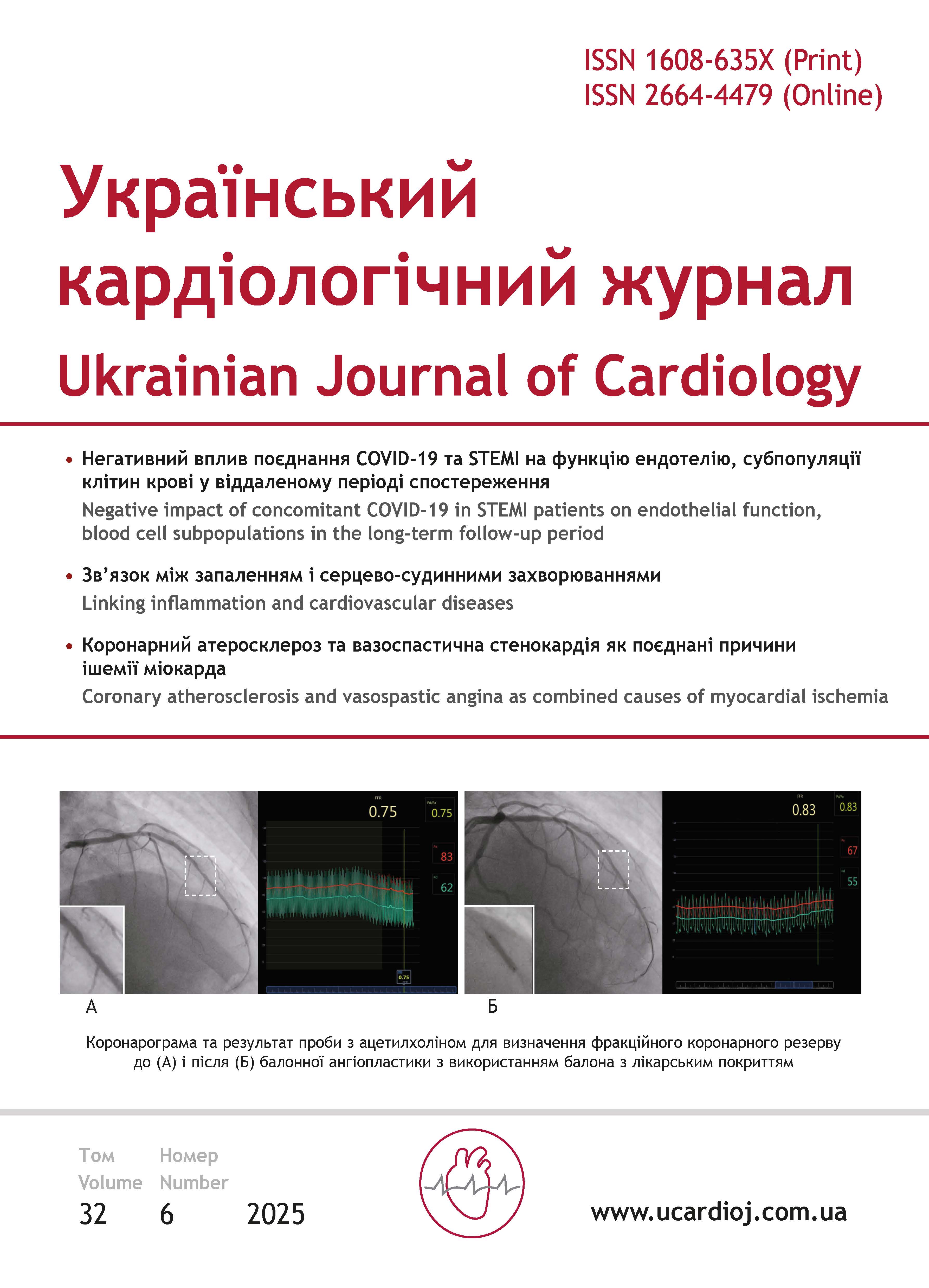
The impact of concomitant COVID-19 in patients with acute myocardial infarction on endothelial function, markers of systemic inflammation, and blood cell subpopulations in the long-term follow-up period
Article Sidebar

This work is licensed under a Creative Commons Attribution-ShareAlike 4.0 International License.
Main Article Content
 https://orcid.org/0000-0002-3563-9627
Abstract
The aim – to assess the course of the coronary artery disease (CAD), the state of endothelial function, intracardiac hemodynamics, and immune system response in the long term follow-up period after ST-elevation myocardial infarction (STEMI) in patients with concomitant COVID-19 for the development of personalized treatment approaches.
Materials and methods. A cohort of patients with CAD (n=60; 85.0 % men; mean age 61.0±1.3 years) was examined 40-48 months after STEMI. Group 1 included patients treated in 2019 before the COVID-19 pandemic (n=30), and group 2 included patients treated during the pandemic in 2020–2022 (n=30) who had documented SARS-CoV-2 infection. The study excluded patients with diabetes mellitus, chronic kidney disease, cancer, chronic inflammatory diseases, high-grade chronic heart failure, and anemia. The examined sample was obtained through screening of 807 patients. All patients underwent a set of clinical, laboratory, and instrumental examinations, which included medical history collection (documented COVID-19 episodes, vaccination history, CAD exacerbations, treatment), cardiac ultrasound, endothelium-dependent vasodilation (EDVD) testing, general laboratory tests, and immunophenotyping of blood cell subpopulations by flow cytometry using the markers CD3, CD4, CD8, CD31, CD34, CD38, and CD309.
Results. Patients in the examined groups did not differ in age or baseline clinical and anamnestic characteristics (including the frequency of documented COVID-19 and vaccination history), and all demonstrated high adherence to treatment. According to EDVD test, endothelial dysfunction was detected in 70 % of patients in both groups, and the mean test values were comparable. Biochemical blood parameters also showed no differences between the groups, nor did inflammatory markers such as C-reactive protein, erythrocyte sedimentation rate, the neutrophil-to-lymphocyte ratio, and the platelet-to-lymphocyte ratio. Correlation analysis of EDVD with inflammatory markers did not reveal any significant associations.
However, immunophenotyping of blood cell subpopulations demonstrated significant differences between the groups. In group 2 patients, who had STEMI with concomitant COVID-19, lower levels of T lymphocytes and higher numbers of immature T-cell were observed, while the levels of T-helpers, T-suppressors, and their ratio did not differ from those in group 1. At the same time, the number of hematopoietic progenitor cells, activated T- and B-lymphocytes, as well as cells expressing vascular endothelial growth factor (VEGF) receptors, were increased. In this group, no significant correlations were found between EDVD results and any clinical, laboratory, or instrumental parameters, nor with the results of blood cell subpopulation phenotyping.
When comparing subgroups of patients with markedly reduced endothelial function (values below the group mean), group 2 showed higher monocyte counts, lower lymphocyte counts, increased populations of activated T- and B-lymphocytes and immature T-lymphocytes, together with elevated levels of repair markers (CD309+ endothelial progenitors), while markers of endothelial activation and damage remained unchanged.
Conclusions. A study conducted in a carefully selected cohort of patients with CAD who had previously experienced STEMI revealed the negative consequences of its combination with COVID-19. Despite similar clinical, instrumental, and laboratory characteristics between the groups, patients with concomitant COVID-19 demonstrated impaired mechanisms of endothelial function regulation and immune system dysfunction, characterized by the development of a pro-inflammatory cellular phenotype. These changes occurred against the background of vascular injury and were accompanied by secondary activation of proangiogenic reparative potential.
Article Details
Keywords:
References
Hacker K. The Burden of Chronic Disease. Mayo Clin Proc Innov Qual Outcomes. 2024 Jan 20;8(1):112-119. https://doi.org/10.1016/j.mayocpiqo.2023.08.005
Rus M, Ardelean AI, Andronie-Cioara FL, Filimon GC. Myocardial Infarction during the COVID-19 Pandemic: Long-Term Outcomes and Prognosis—A Systematic Review. Life (Basel). 2024 Jan 31;14(2):202. https://doi.org/10.3390/life14020202
Ghamar Talepoor A, Doroudchi M. Immunosenescence in atherosclerosis: A role for chronic viral infections. Front Immunol. 2022 Aug 17;13:945016. https://doi.org/10.3389/fimmu.2022.945016. PMID: 36059478; PMCID: PMC9428721
Nanavaty D, Sinha R, Kaul D, Sanghvi A, Kumar V, Vachhani B, Singh S, Devarakonda P, Reddy S, Verghese D. Impact of COVID-19 on Acute Myocardial Infarction: A National Inpatient Sample Analysis. Curr Probl Cardiol. 2024 Jan;49(1 Pt A):102030. https://doi.org/10.1016/j.cpcardiol.2023.102030. Epub 2023 Aug 11. PMID: 37573898
Markson FE, Akuna E, Lim CY, Khemani L, Amanullah A. The impact of COVID-19 on hospitalization outcomes of patients with acute myocardial infarction in the USA. Am Heart J Plus. 2023 Aug; 32:100305. https://doi.org/10.1016/j.ahjo.2023.100305
Basu-Ray I, Almaddah Nk, Vaqar S, et al. Cardiac Manifestations of Coronavirus (COVID-19) [Updated 2024 Feb 12]. In: StatPearls [Internet]. Treasure Island (FL): StatPearls Publishing; 2025 Jan-. Available from: https://www.ncbi.nlm.nih.gov/books/NBK556152/
Hilser JR, Spencer NJ, Afshari K, Gilliland FD, Hu H, Deb A, Lusis AJ, Tang WHW, Hartiala JA, Hazen SL, Allayee H. COVID-19 Is a Coronary Artery Disease Risk Equivalent and Exhibits a Genetic Interaction With ABO Blood Type. Arterioscler Thromb Vasc Biol. 2024 Nov;44(11):2321-2333. https://doi.org/10.1161/ATVBAHA.124.321001
Terlecki M, Wojciechowska W, Klocek M, Olszanecka A, Bednarski A, Drożdż T, Pavlinec C, Lis P, Zając M, Rusinek J, Siudak Z, Bartuś S, Rajzer M. Impact of concomitant COVID-19 on the outcome of patients with acute myocardial infarction undergoing coronary artery angiography. Front Cardiovasc Med. 2022 Sep 22;9:917250. https://doi.org/10.3389/fcvm.2022.917250
Novitalia B, Suryawan IGR, Subagjo A, Firdani M, Intan RE. Endothelial dysfunction by brachial artery flow-mediated dilatation as predictor of major adverse cardiovascular event in acute coronary syndrome. Intern J Health Sciences. 2022; 6(S9): 336-353. https://doi.org/10.53730/ijhs.v6nS9.12263
Galderisi M, Cosyns B, Edvardsen T, Cardim N, Delgado V, et al.; 2016–2018 EACVI Scientific Documents Committee; 2016–2018 EACVI Scientific Documents Committee. Standardization of adult transthoracic echocardiography reporting in agreement with recent chamber quantification, diastolic function, and heart valve disease recommendations: an expert consensus document of the European Association of Cardiovascular Imaging. Eur Heart J Cardiovasc Imaging. 2017 Dec 1;18(12):1301-1310. https://doi.org/10.1093/ehjci/jex244
Corretti M, Anderson T, Benjamin E, et al. Guidelines for the ultrasound assessment of endothelial-dependent flow-mediated vasodilation of the brachial artery: A report of the International Brachial Artery Reactivity Task Force. JACC. 2002 Jan;39(2):257-265. https://doi.org/10.1016/S0735-1097(01)01746-6
Bukreieva T, Kyryk V, Nikulina V, Svitina H, Vega A, Chybisov O, Shablii I, Mankovska O, Lobyntseva G, Nemtinov P, Skrypkina I, Shablii V. Dynamic changes in radiological parameters, immune cells, selected miRNAs, and cytokine levels in peripheral blood of patients with severe COVID‑19. Biomed Rep. 2023 Mar 21;18(5):33. https://doi.org/10.3892/br.2023.1615
Moll-Bernardes R, Camargo GC, Silvestre-Sousa A, Barroso JM, Ferreira JR, Tortelly MB, Pimentel AL, Figueiredo ACBS, Schaustz EB, Secco JCP, et al. Immune Profile and MRI-Detected Cardiac Fibrosis and Edema in Hypertensive and Non-Hypertensive Patients with COVID-19. J Clin Med. 2024;13:7317. https://doi.org/10.3390/jcm13237317
Mancuso P, Gidaro A, Gregato G, Raveane A, Cremonesi P, Quarna J, Caccia S, Gusso L, Rusconi S, Giacomelli A, Cogliati C, Bertolini F. Circulating endothelial progenitors are increased in COVID-19 patients and correlate with SARS-CoV-2 RNA in severe cases. J Thromb Haemost. 2020 Oct; 18(10):2744-2750. https://doi.org/10.1111/jth.15044
Chioh FW, Fong SW, Young BE, Wu KX, Siau A, Krishnan S, Chan YH, Carissimo G, Teo LL, Gao F, Tan RS, Zhong L, Koh AS, Tan SY, Tambyah PA, Renia L, Ng LF, Lye DC, Cheung C. Convalescent COVID-19 patients are susceptible to endothelial dysfunction due to persistent immune activation. eLife, 202110, e64909. https://doi.org/10.7554/eLife.64909
Chang D, Wang J, Zhu S, Shentu Y, Wang Y, Zhu P, Ono M. Cellular senescence and cell therapy in cardiovascular diseases. Stem Cell Res Ther. 2025 Nov 5;16(1):613. https://doi.org/10.1186/s13287-025-04731-6
Coppé JP, Patil CK, Rodier F, Sun Y, Muñoz DP, Goldstein J, Nelson PS, Desprez PY, Campisi J. Senescence-associated secretory phenotypes reveal cell-nonautonomous functions of oncogenic RAS and the p53 tumor suppressor. PLoS Biol. 2008 Dec 2;6(12):2853-68. https://doi.org/10.1371/journal.pbio.0060301
Wiley CD, Campisi J. The metabolic roots of senescence: mechanisms and opportunities for intervention. Nat Metab. 2021 Oct;3(10):1290-1301. https://doi.org/10.1038/s42255-021-00483-8